Weekly Seminar Series
Mondays, 4-5 p.m. | Health Sciences Learning Center
No Seminar December 1
Fall 2025 Seminar Series
This is an accordion element with a series of buttons that open and close related content panels.
December 8 - Marina Emborg, MD, PhD | Cell-based therapies for Parkinson’s disease

Cell-based therapies for Parkinson’s disease
Marina Emborg, MD, PhD
Professor, University of Wisconsin-Madison
Parkinson’s disease (PD) is a progressive neurodegenerative disorder, characterized by the loss of nigral dopaminergic neurons that project into the striatum. Cell-replacement strategies are envisioned as a solution to repair the brain of patients with PD. In this presentation, I will discuss our pioneer work to develop cell-based therapies for PD and the critical role that nonhuman primate models play in the safe clinical translation of 1st-in-class therapies. Lastly, I will discuss our collaboration with Aspen Neurosciences to create an innovative neurosurgical approach for intracerebral delivery of autologous cells, which led to a successful 1/2a clinical trial for PD patients.
November 24 - Steve Peterson, PhD | Prompt Gamma Imaging: Verifying Proton Therapy Treatment Dose

Prompt Gamma Imaging: Verifying Proton Therapy Treatment Dose
Steve Peterson, PhD
Associate Professor and Head of Department, University of Cape Town – Department of Physics
Proton radiation therapy provides exceptional benefits over traditional photon therapy for the treatment of cancer, resulting in lower dose to the patient and less treatment side-effects. This benefit is achieved because the protons deposit most of their energy (dose) directly into the tumor volume. Unfortunately, this concentrated energy deposition makes it difficult to produce accurate pictures of the treatment dose within a patient.
Prompt Gamma Imaging (PGI) is a promising imaging technique for producing in-vivo images of dose delivery during proton radiotherapy. PGI uses solid-state gamma-ray detectors to reconstruct images of the scattered secondary prompt gammas produced during a proton therapy treatment. The talk will discuss the work at UCT with M3D and the University of Maryland Medical Center to develop a clinical dose verification system including a nozzle-mounted PGI system.
Lastly, this talk will also discuss the UCT Proton Therapy Initiative that is working to bring clinical proton therapy to the African continent.
November 17 - Paulina Galavis, PhD | Resilience in Medical Physics: Thriving in a Demanding Profession

Resilience in Medical Physics: Thriving in a Demanding Profession
Paulina E. Galavis, PhD Director of Physics, NYU Langone Health
Medical physics is a high-stakes, rapidly evolving profession that demands precision, adaptability, and sustained focus. Physicists face growing workloads, staffing challenges, regulatory requirements, and constant technological advances, all while ensuring top-quality patient care. Students and trainees encounter additional challenges, including steep learning curves, intense clinical expectations, and uncertainty about future career paths.
The development of resilience at both individual and group levels is crucial for achieving sustained success and enhancing overall well-being. This talk will explore practical strategies for recognizing stress, cultivating adaptability, and fostering supportive environments at personal, team, and departmental levels. The focus will be on implementing effective strategies for stress management, promoting adaptability, encouraging the development of supportive communities, and establishing environments in which physicists, students, and trainees are enabled to thrive rather than merely persevere.
November 10 - Basak E. Dogan, MD | Optoacoustic Breast Imaging: Current status and future trends in clinical application

Optoacoustic Breast Imaging: Current status and future trends in clinical application
Basak E. Dogan, MD
Clinical Professor, Director of Breast Imaging Research
The University of Texas Southwestern Medical Center
Optoacoustic (photoacoustic) breast imaging is an emerging hybrid modality that combines the high spatial resolution of ultrasound with the functional contrast of optical imaging. By detecting ultrasound waves generated from transient thermoelastic expansion after tissue absorption of pulsed laser light, this technique provides non-invasive, radiation-free, and contrast-free assessment of breast tissue. Clinical studies have demonstrated that optoacoustic ultrasound (OA/US) can increase specificity in differentiating benign from malignant breast lesions, significantly reducing unnecessary biopsies. Moreover, semi-quantitative OA/US features correlate with breast cancer molecular subtypes, tumor hypoxia, and angiogenic activity, offering potential for noninvasive biological profiling.
Recent research indicates that OA/US features may predict nodal metastatic burden, highlighting its prognostic value. Limitations include depth penetration, signal variability, and cost. Nevertheless, OA/US represents a promising adjunct to conventional ultrasound, with potential applications in diagnosis, subtype characterization, and treatment response monitoring, including anti-angiogenic and radiotherapy strategies.
November 3 - Eszter Boros, PhD | New Chemical Tools for Next Generation Theranostics
New Chemical Tools for Next Generation Theranostics
Eszter Boros, PhD
Associate Professor of Chemistry
University of Wisconsin–Madison, Department of Chemistry
Radio-theranostics are a rapidly growing area of preclinical and clinical research; the vast majority of theranostic agents utilize radioactive metal ions. My research group specializes in developing bespoke, coordination chemistry approaches to harness validated and emerging radioisotopes. In this seminar, I will discuss 3 case studies of coordination chemistry saving the day in new and unusual ways: 1) a single molecule approach to the chelation of F-18, Ga-68 and Lu-177 2) a metal ion induced, self-degradation strategy to modulate the pharmacokinetics of Ga-68 radiopharmaceuticals, b) a new chemical tool to capture an unchelatable metal ion isotope (Ti-45).
October 27 - Ahtesham Khan, PhD | Electron FLASH Dosimetry: Current Status and Challenges
Electron FLASH Dosimetry: Current Status and Challenges
Ahtesham Khan, PhD
Assistant Professor
University of Wisconsin–Madison
Electron FLASH radiotherapy (eFLASH-RT) has garnered significant interest due to its potential to improve the therapeutic index of radiation treatments. However, accurate dosimetry for pulsed beams at ultra-high dose rates remains one of the primary barriers to clinical translation. This presentation will review the status and key challenges in electron FLASH dosimetry, with an emphasis on both reference and verification dosimetry. The role of ultra-thin parallel plate ionization chambers in establishing reference dosimetry will be discussed, along with the application of beam current transformers for real-time beam monitoring. Complementary use of passive dosimeters for independent dose verification will also be highlighted. Finally, existing limitations and areas requiring further investigation—such as charge build-up effects and standardization of protocols—will be explored. Together, these topics provide an overview of the progress to date and the critical steps needed to enable accurate and reliable dosimetry for eFLASH-RT.
October 20 - Minglei Kang, PhD, DABR, and Shannon O'Reilly, PhD, DABR | Something to Bragg About: Advancements in Proton Therapy Imaging and Planning at UW
Something to Bragg About: Advancements in Proton Therapy Imaging and Planning at UW
Join us for an overview of the development and clinical integration of the new state-of-the-art proton therapy center at UW-Madison which features a gantry room as well as a fixed-beam room with upright CT. We will explore the global landscape of proton therapy, highlight innovations and examine disparities in access. We will discuss advances in imaging and treatment planning, implementation of motion management (surface guidance and real-time gating) and adaptive therapy. This will be followed by ongoing and future research initiatives at the center, with a focus on cutting-edge technologies such as FLASH therapy and the emerging potential of proton arc therapy.

October 13 - Jim Delikatny, PhD | Targeted NIR Fluorescent Dyes for Intraoperative Lung Cancer Visualization
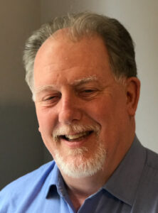
Targeted NIR Fluorescent Dyes for Intraoperative Lung Cancer Visualization
Jim Delikatny, PhD
Professor of Radiology
The University of Pennsylvania
Fluorescence guided surgery (FGS) is an emerging technique employed by oncological surgeons for accurate tumor margin detection, identification of synchronous lesions and locoregional micrometastases, leading to more complete tumor excision and improved patient outcomes. Critical to this effort is the development of near infrared (NIR) fluorescent contrast agents for local or systemic administration that target specific cancer biomarkers. Employing fluorophores that emit in the NIR-I and NIR-II (SWIR) ranges provide greater tissue depth penetration, reduced light scattering and reduced autofluorescence that allow for deeper tissue imaging.
This seminar describes our efforts to design and evaluate targeted and activatable NIR I and II fluorescent imaging probes for the detection of breast and lung cancers in mouse models. We have focused on developing probes that target choline kinase (ChoKa) and cytosolic phospholipase A2 (cPLA2), critical enzymes in lipid anabolism and signaling. We will describe the translation of these probes into veterinary clinical trials for surgical margin detection in spontaneous non-small cell lung tumors in a patient canine population, and our progress towards initiating a human clinical trial.
October 6 - Mario Fabiilli, PhD | The power of bubbles: ultrasound-responsive biomaterials for blood vessel and bone regeneration
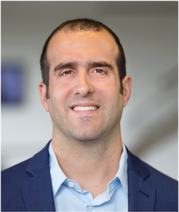
The power of bubbles: ultrasound-responsive biomaterials for blood vessel and bone regeneration
Mario Fabiilli, PhD
Associate Professor of Radiology and Biomedical Engineering
University of Michigan – Ann Arbor
Implantable biomaterials containing bioactive molecules and/or cells can stimulate regeneration of various types of tissue. Regeneration is guided by biochemical and biophysical cues within the biomaterial. However, with conventional biomaterials, cues are preprogrammed when the biomaterial is prepared and thus cannot be dynamically changed after implantation. This limits both fundamental and translational studies, including the personalization of regenerative therapies. We are developing biomaterials that can be modulated non-invasively and in an on-demand manner using focused ultrasound. One strategy we employ is creating composite hydrogels with a phase-shift emulsion, which are liquid droplets that vaporize into bubbles upon exposure to ultrasound. By leveraging the unique interactions between ultrasound and phase-shift emulsions, we have developed strategies for controlling the release of bioactive factors as well as modulating the microarchitecture and physical properties of hydrogels. This talk will highlight our work in revascularizing ischemic tissue by promoting blood vessel formation and stimulating bone growth by enhancing osteogenic differentiation of mesenchymal stromal cells.
September 29 - Cameron Symposium Guest Muyinatu “Bisi” Bell, PhD | Ultrasound and Photoacoustic Imaging

Ultrasound and Photoacoustic Imaging
Muyinatu “Bisi” Bell, PhD
John C. Malone Associate Professor
Johns Hopkins University
Ultrasound and photoacoustic imaging are two non-invasive techniques that allow us to peek into the body to visualize internal anatomy, offering advantageous diagnostic, surgical, and interventional guidance information. Ultrasound imaging transmits sound that is reflected and detected by an array of sensors placed in contact with the skin. Photoacoustic imaging transmits light that is absorbed, causing thermal expansion which generates sound that can be detected with the same senor array. In both cases, the sensed signals are processed using beamformers to display image information. However, conventional beamformers exclusively rely on signal amplitudes, ignore the impact of light transmission through darker skin tones, or assume uniform properties (e.g., sound speed) which overlook naturally occurring intra- and inter-patient variations.
In this talk, I will provide real-world examples of the medical imaging inequities that result from conventional beamformer design choices. I will then describe techniques to address these inequities using signal processing innovations that consider spatial correlations rather than signal amplitudes. Specific clinical applications that have the greatest potential to benefit from a coherence-based imaging approach include cardiovascular health assessments, breast cancer diagnosis and treatment, biopsies, neurosurgery, teleoperated robotic surgery, and wearable health applications with flexible arrays.
September 22 - Filiz Yesilköy, PhD | Mid-infrared imaging and sensing in biomedicine

Mid-infrared imaging and sensing in biomedicine
Filiz Yesilköy, PhD
Vilas and Grainger Assistant Professor
University of Wisconsin–Madison
Label-free vibrational spectroscopy in the mid-infrared (mid-IR) spectrum (𝜆 = 2.5–25 µm) is a powerful tool for noninvasive analysis of biological samples without lengthy sample staining and preparation. This technique enables the detection of biomolecules based on their unique spectral fingerprints, where molecular vibrational energy states are inscribed as absorption peaks. Spectrochemical fingerprinting is particularly promising for biomedical diagnostics, disease monitoring, and biomarker discovery because it can simultaneously capture diverse molecular structures from biospecimens. However, when analyzing complex biological samples, conventional mid-IR absorption spectroscopy faces significant challenges. Specifically, heterogeneity in sample thickness and composition and optical scattering distort and congest spectral data, impeding reliable chemometric analyses. My work focuses on developing advanced optical analytical platforms by mastering nanotechnology, plasmonics, and metasurfaces to address the major challenges of mid-IR chemical analysis. In my talk, I will introduce numerous photonic metasurface technologies and highlight the unique light-matter interactions that are key for developing advanced biochemical sensors for medical applications.
September 15 - Chris Flask, PhD | Magnetic Resonance Fingerprinting: Opportunities and Challenges for Both Basic Science and Human Imaging
Magnetic Resonance Fingerprinting: Opportunities and Challenges for Both Basic Science and Human Imaging
Chris Flask, PhD
Professor, Department of Radiology
Case Western Reserve University
Magnetic Resonance Fingerprinting (MRF) was first developed in 2013 to rapidly generate quantitative T1 and T2 relaxation time maps. This presentation will introduce the primary components of MRF including: highly-undersampled k-space trajectories, intentional variation in the acquisition parameters, MRF dictionary variation, and the parameter matching process. This talk will discuss how these different components can be combined and optimized in a highly rationalized in order to meet the demands for specific imaging applications. First, I will discuss how MRF methods are designed for human body imaging applications, then I will discuss the challenges and opportunities for implementing MRF on high field MRI scanners for preclinical imaging studies. In particular, I will discuss my ongoing collaborations with Dr. Marty Pagel in the development of Dynamic Contrast Enhanced – MRF methods to assess tumor perfusion in breast cancer mouse models.
September 8 - Edmond Sterpin, PhD | Online adaptive proton therapy and the central role of time

Online adaptive proton therapy and the central role of time
Edmond Sterpin, PhD
Professor, KU Leuven & UCLouvain
Recent developments in online adaptive proton therapy (OAPT) have focused on overcoming anatomical variations during treatment through fast, automated workflows that integrate imaging, contour adaptation, plan re-optimization, and quality assurance. Advances include automated dose-restoration strategies for maintaining target coverage and organ-at-risk protection, as well as collaborative efforts to streamline near-real-time adaptation including full image segmentation supported by artificial intelligence. Recent investigations have highlighted that while rapid adaptation can provide substantial dosimetric and predicted toxicity benefits from an individual patient point of view, these gains decrease sharply at the level of the patient population when adaptation becomes time-intensive, emphasizing the need for highly efficient clinical workflows. Together, these findings reinforce that both plan quality and adaptation speed are critical for translating OAPT into routine clinical practice.
Spring 2025 Seminar Series
This is an accordion element with a series of buttons that open and close related content panels.
April 28 - Shreya Goel, PhD | Photoacoustic Imaging to Assess Oncologic Treatment Response and Toxicity
 Photoacoustic Imaging to Assess Oncologic Treatment Response and Toxicity
Shreya Goel, PhD
Assistant Professor of Molecular Pharmaceutics, University of Utah
Photoacoustic Imaging (PAI) combines optical imaging with ultrasound to provide noninvasive, multiparametric, high-resolution assessment of deep tissues. Compared to conventional imaging modalities that require ionizing radiation, PAI can provide distinct advantages for vulnerable patient populations. In the first part of this talk, I will discuss the utility of oxygen-enhanced (OE) PAI for treatment monitoring in mouse models of pediatric rhabdoid tumors. We show that OE-PAI can visualize spatiotemporal dynamics of hypoxia modulation by repurposed metabolic inhibitors, that can be used to optimize combination treatment with low-dose radiotherapy.
In the second part, I will describe our ongoing efforts to evaluate skin toxicities (hand-foot syndrome, HFS) that are frequently observed in patients undergoing cancer chemotherapy. We utilize contrast-enhanced-PAI to quantitatively visualize primary drug-induced skin permeability in mouse model of HFS, and its relation to secondary hyperpermeability resulting in massive drug accumulation and tissue damage.
April 21 - Christian Farrar, PhD | Molecular Imaging of Oncolytic Virotherapy with CEST Magnetic Resonance Fingerprinting and CEST Reporter Genes

Molecular Imaging of Oncolytic Virotherapy with CEST Magnetic Resonance Fingerprinting and CEST Reporter Genes
Christian Farrar, PhD
Associate Professor of Radiology, Martinos Center for Biomedical Imaging, Department of Radiology, Massachusetts General Hospital and Harvard Medical School
Cell- and viral-based therapeutics hold great promise for revolutionizing the treatment of many diseases. However, optimizing such biological therapies and assessing their efficacy depends on the ability to both monitor the spread and persistence of the therapeutic agent and evaluate the tissue molecular response. Our work has been focused on developing improved molecular imaging tools for guiding oncolytic virotherapy treatment of glioblastoma. We have recently pioneered a quantitative magnetic resonance fingerprinting (MRF) based chemical exchange saturation transfer (CEST) method to both monitor tumor apoptotic response to oncolytic virotherapy, characterized by decreased protein synthesis and intracellular pH, as well as to track the delivery and spread of the oncolytic virus using a CEST reporter gene. Quantitative CEST-MRF, with its sensitivity to pH and protein and metabolite concentrations, provides a powerful tool for non-invasive, contrast-agent free, molecular imaging.
April 14 - Randy Bartels, PhD | Computational adaptive optics nonlinear holographic microscopy for tissue imaging
Computational adaptive optics nonlinear holographic microscopy for tissue imaging
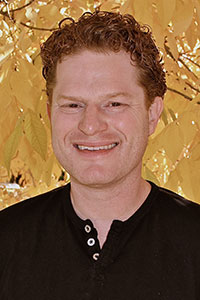 Randy Bartels, PhD
Principal Investigator at the Morgridge Institute for Research and Professor of Biomedical Engineering, UW-Madison
Optical microscopy plays a pivotal role in the understanding of spatial and temporal dynamics of biological systems and for probing material systems. Light interacts gently with biological systems, which makes imaging extremely powerful for observing living systems. As a result, optical microscopy enables everything from the discovery of basic biological processes to the ability to diagnose disease to the discovery of new materials. Optical microscopy is primarily bound by three limits: spatial resolution, molecular specificity of imaging targets, and imaging depth in tissue. My group develops new methods for extracting a greater range of information from optical microscopy.
I will discuss several aspects of computational imaging with widefield nonlinear microscopy using second harmonic generation (SHG), third harmonic generation (THG), and coherent anti-Stokes Raman scattering (CARS). These imaging methods are extremely useful for imaging studies ranging from biological samples to novel material systems. In biological samples, SHG, THG, and CARS signal generation is dominated by signals from collagen in the extracellular matrix and from muscle fibers. I will discuss computational adaptive optics for SHG and THG holographic imaging. In addition, I will discuss a new widefield computational super resolution CARS microscopy that offers a powerful new approach to vibrational spectroscopic imaging.
April 7 - Jordan Perchik, MD | I’m the Problem: How to respond when you have committed a microaggression
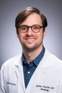
I’m the Problem: How to respond when you have committed a microaggression
Jordan Perchik, MD
Assistant Professor
Department of Radiology
University of Alabama-Birmingham
A welcoming work environment is a critical component to a successful and collaborative workforce. In an ever-diversifying healthcare field, intentional steps must be taken to recognize and effectively intervene when microaggressions occur. It is also important to recognize that microaggressions, or subtle acts of exclusion, are often unintentional, and sometimes, we may commit microaggressions, ourselves. In this presentation, we will discuss what microaggressions are, how to recognize and respond to microaggressions, and how to respond and learn from mistakes when you have committed a microaggression.
March 31 (no seminar March 24) - Jeff Squier, PhD | Structured light imaging: optical metrology for applications from advanced manufacturing to the neurosciences
Structured light imaging: optical metrology for applications from advanced manufacturing to the neurosciences
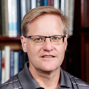 Jeff Squier, PhD
Professor, Department of Physics
Colorado School of Mines
Structured illumination imaging enables the use of extended excitation sources for use in environments where the signal light can be scattered along the collection path toward the detector. Like point excitation, structured light extended excitation source imaging is compatible with single element detection, hence its utility in imaging in specimens that result in scattered signal light. Further, the structure makes extended excitation geometries like a line excitation compatible with point geometries in terms of resolution, and in fact these extended geometries can possess broader spatial frequency support. The system is also image contrast agnostic resulting in enhanced spatial frequency support across mechanisms: linear and nonlinear excitation, coherent and incoherent signal generation. We have developed structured illumination imaging systems with exposures times ranging from 100 µs, down to the limit of a single excitation pulse: 30 fs! Results from all these systems will be presented with examples of how these systems can be used to address challenges in advanced manufacturing to the biosciences. We will also show results across contrast mechanisms and signal sources (coherent and incoherent).
March 17 - Daniel Ennis, PhD | Using MRI To Estimate Cardiac Structure and Function

Using MRI To Estimate Cardiac Structure and Function
Daniel Ennis, PhD
Professor of Radiology and Bioengineering (by courtesy)
Stanford University
Magnetic resonance imaging (MRI) is a remarkable technology capable of probing the heart’s structure and function. The goal of these methods is to better understand cardiac performance in health and disease. Diffusion-based MRI methods have been used for decades to explore the microstructural organization of the brain and, more recently, related methods have undergone significant development to enable exploring the mesostructural organization of the heart. This talk will provide an overview of the challenges and solutions to enabling diffusion-based MRI of the heart. Estimates of cardiac mesostructure can then be combined with estimates of cardiac motion to better understand the structure-function relationships and provide insights that go beyond conventional measures of heart function.
March 10 - Paul Harari, MD & Carri Glide-Hurst, PhD | Standing Tall to Cancer: Physics and Physician Highlights of the World’s First Academic Upright Radiation Therapy Program
Standing Tall to Cancer: Physics and Physician Highlights of the World’s First Academic Upright Radiation Therapy Program
 Dr. Paul Harari, MD, FASTRO
Jack Fowler Professor
Department of Human Oncology
Principal Investigator, Wisconsin Head and Neck SPORE Grant
University of Wisconsin School of Medicine and Public Health
 Dr. Carri Glide-Hurst, PhD, DABR, FAAPM
Bhudatt Paliwal Endowed Professor
Associate Chair, Radiation Oncology Physics
Departments of Human Oncology and Medical Physics
University of Wisconsin School of Medicine and Public Health
More than half of cancer patients will receive radiation therapy during their treatment. Historically, radiation therapy has been administered with patients lying down in the supine position with a large gantry rotating around them to treat their cancers from various angles. However, imaging and treating cancer patients in an upright position offers many advantages compared to supine. University of Wisconsin will be pioneering upright positioning for cancer patient treatments through a research installment in WIMR and using a fixed proton radiation beam configuration at the UW Health Eastpark Medical Campus. This talk will feature the journey to bring upright proton treatments to UW from the initial planning to the current stage. A physician and physicist pairing will share the exciting vision and first results with this state of the art system that will enable patients to stand tall against cancer in Wisconsin and beyond.
March 3 - Michael Veronesi, MD, PhD | Brain tumor theranostics: From bedside to bench and back for personalized medicine
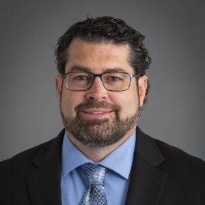
Brain tumor theranostics: From bedside to bench and back for personalized medicine
Michael Veronesi, MD, PhD
Neuroradiologist and Director of Neuro-oncological Imaging
University of Wisconsin–Madison, Department of Radiology
Radionuclide theranostics is showing great promise for personalized medicine, whereby, a patient’s candidacy is first evaluated with molecular imaging to determine the optimal therapy tailored to the individual. Receptor specific therapy response assessment in radionuclide theranostics with follow up molecular imaging allows loop closure in initial patient assessment and next steps in management. Brain tumor theranostics is one area with great potential for personalized theranostics applications. The first part of the talk will introduce clinical, near clinical or clinical research investigational brain tumor PET/MR examples of molecular imaging being performed at the University of Wisconsin. The latter part of the talk will focus in on amino acid theranostics, to include 18F-flouroethyltyrosine (18F-FET) PET imaging and its partner beta emission theranostics compound, 131I-Iodophenylalanine (131I-IPA), both as a clinical trial agent and as part of a preclinical research project at UW in a GBM rodent model. An important theme will also tie in the importance of partnering with industry for optimizing the theranostics approach.
February 24 - Kathleen M. Schmainda, PhD | Harnessing Quantitative Perfusion MRI Metrics for Brain Tumor Treatment Management
 Harnessing Quantitative Perfusion MRI Metrics for Brain Tumor Treatment Management
Kathleen M. Schmainda, PhD
Professor of Biophysics & Radiology
Medical College of Wisconsin
Treated glioblastoma and non-tumor/treated tissue can appear the same on conventional post-contrast MRI, a primary challenge for glioblastoma treatment management today. As a solution, diagnostic biopsy samples from glioblastoma patients were spatially matched to maps of quantitative, standardized relative cerebral blood volume (sRCBV) and thresholds determined to distinguish tumor from non-tumor tissue within contrast enhancement. The regions of true enhancement can be objectively identified using another quantitative method, referred to as deltaT1. The dT1 together with sRCBV are used to generate maps of fractional tumor burden (FTB) that visually display regions of tumor or non-tumor (treatment effect) within regions of contrast enhancement. These methods have demonstrated clinical utility for guiding surgical biopsy, objectively identifying the true extent of resection or ablation, and providing critical information to accurately characterize treatment response to both standard of care and new therapies.
February 17 - John Garrett, PhD, DABR | Value Added CT with Opportunistic Screening: Automated biomarkers, Applications, and Future Directions
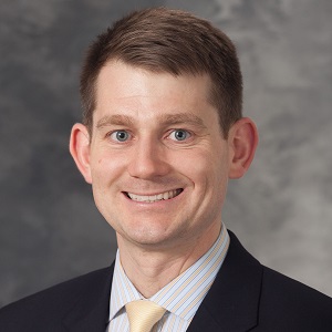 Value Added CT with Opportunistic Screening: Automated biomarkers, Applications, and Future Directions
John Garrett, PhD, DABR
Associate Professor (CHS) of Radiology, Medical Physics, and Biostatistics and Medical Informatics; Director of Imaging Informatics
University of Wisconsin–Madison
Computed Tomography (CT) is a cornerstone of diagnostic imaging, providing critical anatomical and pathological insights. Beyond its diagnostic value, CT also offers untapped potential for opportunistic screening, leveraging existing scans to extract quantitative biomarkers that inform patient health. This presentation explores the concept of value added CT, highlighting how automated analysis tools can unlock this latent value.
We will review state-of-the-art automated biomarkers derived from CT, including body composition analysis, vascular calcifications, and bone density, discussing their implications for screening and risk stratification in chronic diseases like cardiovascular disease, osteoporosis, and cancers. We will explore the impact of CT scan protocol variability and will also discuss the concept of biological aging and how imaging markers might help shed some light on that. Additional case studies will illustrate real-world applications, focusing on technical methods, clinical integration, and the benefits of opportunistic analysis for population health.
Finally, we will discuss future directions, including emerging AI-driven techniques, extension to other modalities, and integration with electronic health records. By repurposing routine CT imaging data, we can enhance the value of radiological studies, driving innovation in preventive care and multidisciplinary collaboration.
February 10 - Jacob Scott, MD, DPhil | Beyond constraint satisfaction: how genomics can usher us into true radiation plan optimization

Beyond constraint satisfaction: how genomics can usher us into true radiation plan optimization
Jacob Scott, MD, DPhil
Professor of Medicine and Physics, Staff Physician-Scientist and Radiation Oncologist
The Cleveland Clinic and Case Western Reserve University
While we say we are making optimal plans for patients in radiation oncology, what we are doing is actually choosing from a number of plans which satisfy constraints. In the absence of continuous functions of outcome, this is the best we can do. However, with the advent of functions relating physics radiation dose to tumor genomics, these continuous functions do exist, opening the door for true multicriteria optimization. In this lecture I will go through the derivation of these continuous functions from tumor genomics using canonical equations of radiation biology, and review the data supporting their mapping to outcome.
February 3 - Summer Gibbs, PhD | Can Near Infrared Nerve-Specific Imaging Improve Surgical Outcomes?
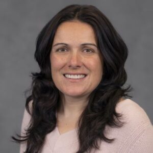
Can Near Infrared Nerve-Specific Imaging Improve Surgical Outcomes?
Summer Gibbs, PhD
Douglas Strain Endowed Professor of Biomedical Engineering
Oregon Health & Sciences University
Fluorescence guided surgery (FGS) is a nascent field, however with ~15,000 clinical FGS system distributed worldwide, its potential to specifically highlight tissues to be resected (e.g., cancer) and avoided (e.g., nerves) has been recognized and there are >125 ongoing clinical trials with novel contrast agents to leverage this clinical imaging technology. Iatrogenic nerve damage is arguably one of the most feared surgical complications as nerve injury is often permanent leaving patients with pain, loss of function and disability. Near infrared (NIR) nerve-specific contrast agent(s) that are spectrally matched to the existing clinical FGS infrastructure have a direct path to clinical translation with broad surgical applicability. However, development of NIR nerve-specific probes has been a substantial challenge as these probes must be small enough to cross the tight blood nerve barrier, but have a sufficient degree of conjugation (i.e., double bounds, which by definition increase the molecular weight) to reach NIR wavelengths. Through a directed fluorophore medicinal chemistry approach, we have designed and developed first-in-kind, small molecule NIR nerve-specific fluorophores. Our team is currently working towards clinical translation of our novel probes as we explore the utility of nerve imaging for a variety of surgical indications including prostatectomy, neurosurgery, endocrine surgeries, head and neck surgeries and orthopedic indications.
January 27 - Ivan Rosado Mendez, PhD | Expanding Horizons in Diagnostic Ultrasound: New Modalities and Their Clinical Impact
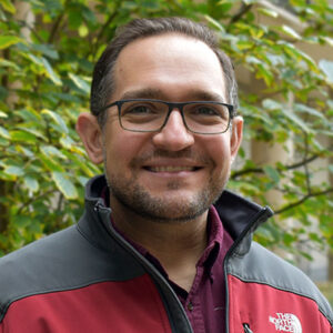
Expanding Horizons in Diagnostic Ultrasound: New Modalities and Their Clinical Impact
Ivan Rosado Mendez, PhD
Assistant Professor, Departments of Medical Physics and Radiology
University of Wisconsin–Madison
Diagnostic ultrasound is at a pivotal juncture. The increasing computational power of ultrasound scanners has enabled the development of novel imaging modalities, revealing new sources of contrast that enhance clinicians’ understanding of disease onset, progression, and response to therapy. Concurrently, the advent of handheld and wearable transducers is democratizing access to diagnostic tools, thereby promoting more equitable access to medical imaging. These advancements present new challenges for medical physicists.
This talk will provide an overview of the biological and clinical motivations behind the development of new ultrasound imaging techniques that uncover novel sources of contrast in diagnostic ultrasound. Emphasis will be placed on their physical foundations and the technical aspects of their implementation. Specific applications from the Quantitative Ultrasound Lab will be presented. The talk will conclude with a critical discussion on the challenges these new techniques face and on potential research avenues to address these challenges and facilitate clinical adoption.
