Medical Physics Seminar – Monday, February 16, 2015
Quantitative pulmonary perfusion using dynamic contrast-enhanced MRI

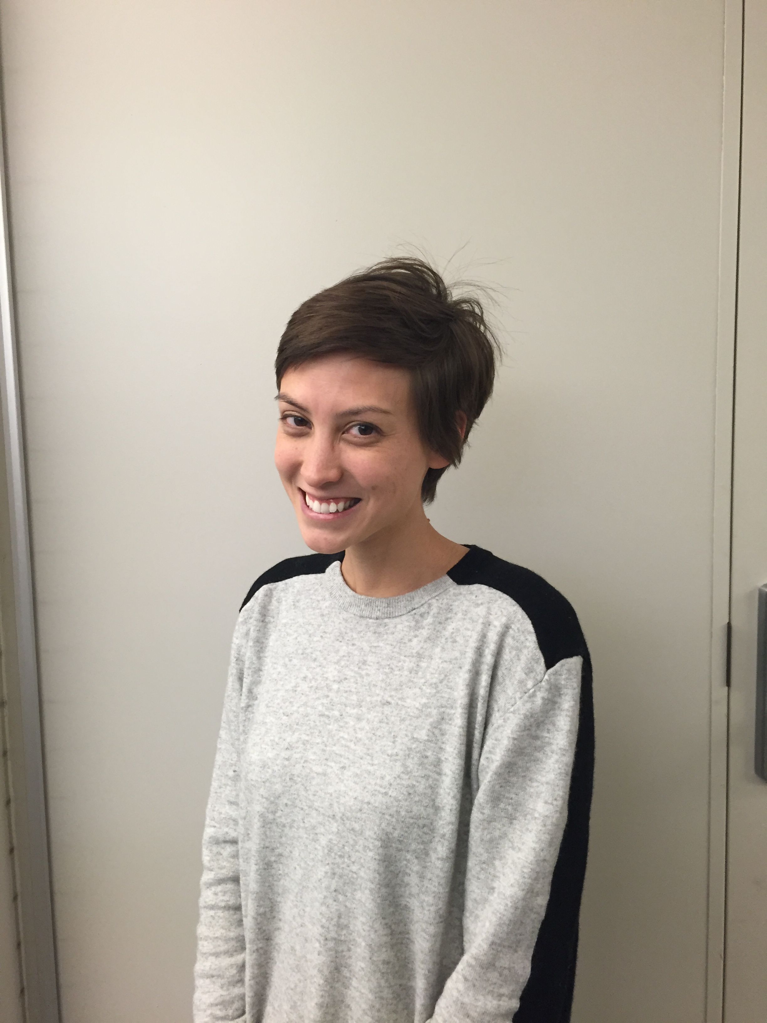
Laura Bell (student of Dr. Thomas Grist)
Research Assistant, Medical Radiation Research Center, Dept of Medical Physics, UW-School of Medicine & Public Health, Madison, WI - USA
Pulmonary perfusion is the process of arterial blood flow through the lung's capillary system [mL/min/g]. Pulmonary perfusion is altered in different lung diseases and during various stages of lung disease making it a potential marker for disease diagnosis, progression, and response to therapy. Magnetic resonance imaging (MRI) has shown promise as a cross-sectional modality capable of imaging both lung function (perfusion) and structure without ionizing radiation. Dynamic contrast-enhanced MRI, also known as "first pass" or "bolus tracking" is commonly implemented to visualize perfusion. It is based on measuring the signal change over time due to the T1-shortening effects of a gadolinium contrast agent. However, several challenges exist with current scanning protocols and post-processing methods to derive hemodynamic parameters. Additionally, imaging of lung structure is typically desired to complement perfusion for complete diagnosis. Current MRI protocols involve two separate scans each optimized for lung structure and function. During this seminar, I will talk about optimizing scanning protocols and post-processing methods for quantitation, and will introduce a technique to simultaneously acquire both lung structure and function in one scan.