Medical Physics Seminar – Monday, February 12, 2018
Ultrasound Transducer Uniformity and Sensitivity Assessment, and Initial Solution to Some CT QA Problems

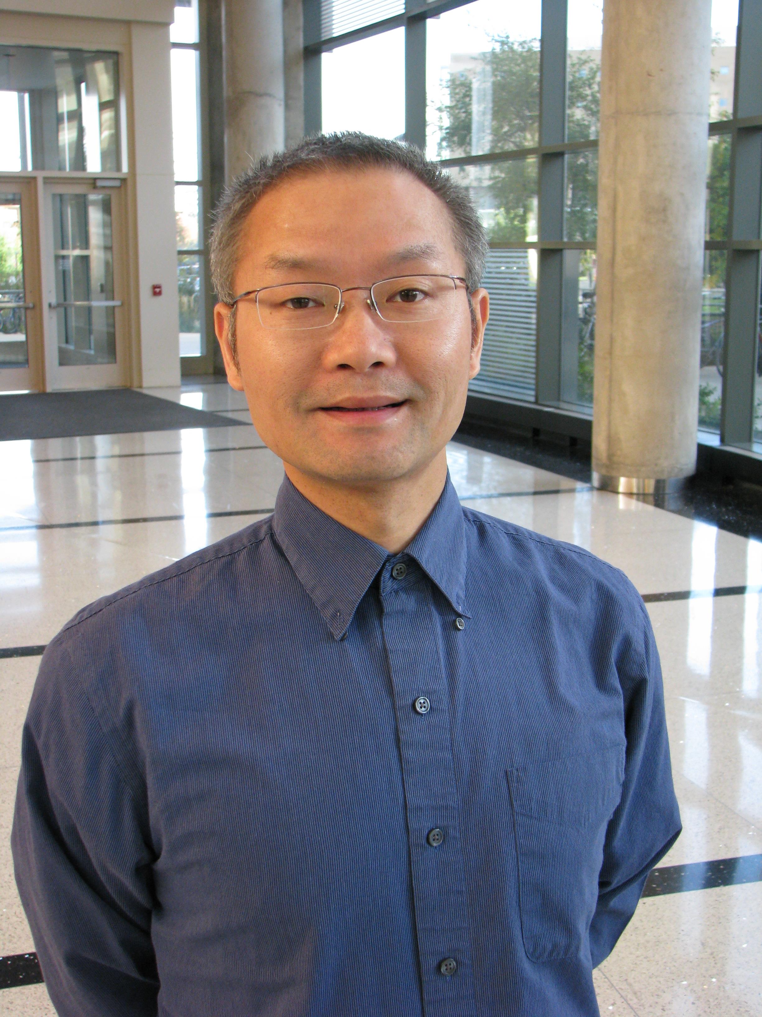
Zhimin Li, PhD (Resident, advised by Dr. Frank N. Ranallo)
Imaging Residency Program, Department of Medical Physics, University of Wisconsin, Madison, WI - USA
Accurate quality assurance (QA) for diagnostic imaging modalities can benefit tremendously from a systematic understanding of various factors that affect imaging characteristics, and from the development of analysis tools for im-proving quantification of such characteristics. In this talk I will discuss my ex-perience in improving QA for two important modalities, Ultrasound and CT. For the Ultrasound part, I will demonstrate how to better assess the depth of penetration (DOP), which is important for evaluating ultrasound system sensi-tivity, by utilizing objective measures of signal to noise. The dependence of DOP on machine settings and phantom variation will be discussed. Then I will discuss a new computerized analysis method that can perform Ultrasound uniformity assessment for the detection of transducer defects. For the CT part, I will discuss phantom design characteristics that can affect the accuracy of measurement of CT numbers of test materials. Automatic analysis of some routine CT QA tasks also will be presented.
An Introduction to Imaging Workflow Analytics and Application to CT Time Efficiency Evaluation in Acute Stroke Response

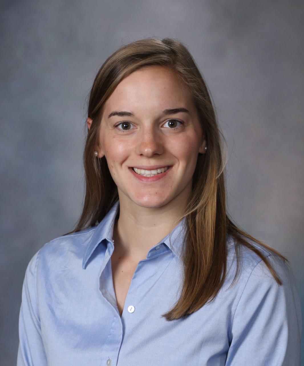
Christina Brunnquell, PhD (Resident, advised by Dr. Frank N. Ranallo)
Imaging Residency Program, Department of Medical Physics, University of Wisconsin, Madison, WI - USA
A wealth of data is associated with each clinical image we gather, including order details in HL7 messaging, clinical context in the electronic health record, and DICOM headers containing information pertaining to the equipment, clini-cal setting, and timing of imaging workflow steps. When collected, sorted, and analyzed intelligently, this data can be used to inform radiology workflow, practices, and training for increased efficiency and value. This talk will intro-duce the role of imaging analytics, the potential role of the imaging physicist in developing and using imaging analytics tools, and the sources of relevant data in the radiology department. It will describe in detail the application of imaging analytics to measuring and improving efficiency of stroke response in CT imag-ing. We retrospectively analyzed CT imaging speed in stroke response to identi-fy institutional trends and opportunities for improvement by extracting time stamps related to image acquisition, reconstruction, and PACS availability from nearly 500 acute stroke CT exams over 1.5 years. We measured statistically and clinically significant effects of scanner location, time of day and year, perfusion processing technique, and performing technologist on imaging efficiency in this time-sensitive setting. This method of monitoring acute stroke imaging perfor-mance facilitates directed training, purchasing justification, and quality im-provement in the acute stroke imaging workflow.