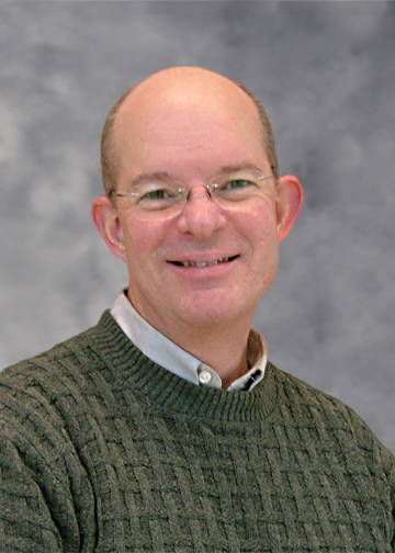
Timothy J. Hall
Professor of Medical Physics
Family?
I have a 70 lb. mutt dog named Blue. It’s just Blue and me. Other than that I have 3 siblings, and my mom is alive and lives in Michigan.
What book is on your nightstand?
Ha! Really? Like I can read at night after reading all day? There is a relatively decent book on the disaster in Fukushima that if I have time to read when I go diving, I might read that.
What accomplishment are you most proud of?
That’s a tough question. I would say training successful students is right up there at the top of the list.
What other career could you see yourself in?
I have always enjoyed economics and could see myself working in that field. Or perhaps diving. I’ve had a passion for diving since I was young and right now that sounds pretty good too.
Hobbies?
I like a wide variety of things. Scuba diving comes readily to mind. I enjoy time with friends, music, cars, landscaping (before I had Blue – he has different ideas of the value of plants), maintaining/improving my house. I also like water sports (fishing, canoeing, kayaking, paddleboarding, etc.), hiking, and biking.
In the Spotlight
What attracted you to the field of Medical Physics?
The main thing that attracted me to medical physics is that there are lots of interesting problems to solve. I first came to UW-Madison for a research opportunity as an undergrad. I spent a summer working with one of my professors who had been working with John Cameron. While I was here I saw all of the job opportunities that were available in Medical Physics and I was impressed because at the time I had been considering high energy physics (where the job market was not that great). That got the ball rolling.
What is your current project, and what do you hope it will accomplish?
We use ultrasound to describe collagen microstructure of tissues. We can easily sense things like how soft/stiff tissues are and can see local variations in stiffness. We can also sense a variety of other properties that describe the tissue microstructure (like how organized it is; whether properties are homogeneous, isotropic, anisotropic, periodically spaced, etc.). From information like this, we hope to reduce the need for serial biopsy to track tissue changes with disease progression or treatment. There might also be situations where this information is sufficient to avoid biopsy or even be used for disease detection and classification. There are two main projects in our research group related to this. One project is targeted toward breast [cancer] imaging with ultrasound, with the intent to improve the specificity of breast ultrasound – especially in women with mammographically dense breasts. Breast ultrasound currently leads to an increased rate of benign biopsy, so improving specificity would hopefully reduce that rate. The other project is in cervical assessment for preterm birth. Ultrasound seems to be able to quantify changes in the cervix over time, and we are hoping to be able to use that to predict women at high risk for pre-term birth.
What are the challenges of working in Medical Physics? What can students learn from these challenges?
Our work is multiscale, highly multidisciplinary and translational. An environment like that brings together a very diverse group of investigators with disparate viewpoints and sometimes very different terminology. More challenging is the very different skill sets among the group of senior investigators. The amusing part, sometimes, is when we discover that we’re using the same terms but mean very different things and use very different assumptions. This creates a great environment for students and postdocs to get exposure to a wide range of research activities and learn to effectively communicate with a wide variety of colleagues.
What’s the best advice you’ve ever received? What advice would you give to students?
Never assume too much. Test your assumptions and validate your programming in as many ways as you can think of. It will save you time in the long run.
What do you think is the future of MRI medical physics?
Interesting question. My guess is that MRI will remain in “developed countries” and only in areas with a large enough population base to justify the cost of implementation and maintenance. So, it will remain rare in areas with low population density. I also think that the integration of PET with MRI will continue to expand its use and what can be learned from it, but those systems will be even more rare.
I think the future of medical physics will be a continued diversification, just like that seen in physics, engineering and even subspecialties like electrical engineering. I think the horizons are broadening and much more effort will continue to go into ‘putting pieces together’ for a better understanding all that we do in image science, diagnostics and therapeutics.
If you were awarded a million-dollar research grant, what would you do with it?
There are a lot of things I could do with the money. The main thing is funding students to further their investigations, and being able to purchase equipment that would get optical imaging more ingrained in WIMR. However, I’ve had a few ‘large’ grants and amazingly a million dollars doesn’t go very far.

