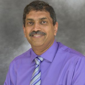
Tomy Varghese
Credentials: PhD
Position title: Professor, Medical Physics
Co-Vice Chair for Graduate Program
Email: tvarghese@wisc.edu
Phone: 608/262-2413
Address:
1159 Wisconsin Institutes for Medical Research
UW-Medical Physics, Ultrasound Imaging
Madison, WI 53705

My primary research interests are in signal, image processing and machine learning applications in medical ultrasound imaging for the early detection of cancer. Early detection of cancers can significantly improve patient survival rate and lead to effective treatment. Aims of our research are to develop signal-processing and machine learning approaches for extracting relevant tissue microstructure information embedded in the spectra of RF broadband ultrasonic echoes. The ultimate goal is to provide fast, reliable and well-understood signal processing techniques for the early detection of malignant tissue using ultrasound. A unique aspect of this research is its focus on characterizing Fourier phase and magnitude of RF ultrasonic echoes with spectral redundancy characterized by the spectral autocorrelation function (also called the Generalized Spectrum). Disruptions in the normal structure of liver and breast tissue due to invading cancers, are detected by examining the changes in the quasi-periodic nature of the liver tissue and the presence of specular scatterers (calcifications in malignant tissue) in breast tissue using this innovative approach. This method is more accurate than the traditional techniques used to estimate the scatterer spacing.
My current research deals with the development of Elastography. Elastography is rapidly developing into a new ultrasonic imaging modality, capable of producing images of internal strain or tissue elasticity. Elastography is a method for imaging the elastic properties of compliant tissues, producing gray scale strain images referred to as elastograms. You can think of Elastography as a “high tech” form of palpation. Elastography has been used for imaging and characterizing tumors in the breast, prostate, kidney, liver, muscle and other tissues. Ultrasonic visualization is widely used in practically all-medical specialties. The proposed technique could therefore have a large impact on medical practice in the United States. Elastography has also been implemented using MRI, Optical Coherence Tomography (OCT) and other imaging modalities.
Education
B.E., (1988) Instrumentation Techology University of Mysore, SJCE, Mysore, India
M.S., (1992) Electrical Engineering, University of Kentucky, Lexington, KY
Ph.D., (1995) Electrical Engineering, University of Kentucky, Lexington, KY
Department Affilations
Medical Physics, University of Wisconsin School of Medicine and Public Health
Biomedical Engineering, University of Wisconsin-Madison
Electrical and Computer Engineering, University of Wisconsin-Madison
Positions
- 07/1995-07/2000, Post-Doctoral Fellow, Radiology, University of Texas Medical School, Houston
- 08/2000 – 04/2006, Assistant Professor, University of Wisconsin, Madison
- 04/2006 – 03/2011, Associate Professor, University of Wisconsin, Madison
- 03/2011 – Present, Professor, University of Wisconsin, Madison
Research Interests
- Elastography
- Clinical Strain and Shear wave imaging
- Signal, Image Processing and Machine Learning Applications in Ultrasound
- Quantitative Ultrasound Imaging
- Pre-clinical Small Animal Photoacoustic and High-Frequency Ultrasound
Awards and Honors
- Second prize, 2005 Endourological Society Basic Science Essay Competition: Gyan Pareek, ER. Wilkinson, S Bharat, Tomy Varghese, PF. Laeseke, FT. Lee Jr., TF. Warner, JA. Zagzebski, SY. Nakada, Elastographic Measurements of in-vivo Radiofrequency Ablation Lesions of the Kidney, Journal of Endourology Nov 2006, Vol. 20, No. 11: 959-964.
- Sigma Xi Research Day Best Poster Award: J. Ophir, EE Konofagou, T. Varghese, F. Kallel, S. K. Alam, and I. Cespedes, “Elastography: A quantitative method for imaging the elasticity of biological tissue”, April 3, University of Houston, (1997).
- Member, ETA Kappa Nu (Electrical Engineering Honor Society)
- Outstanding Teacher evaluation, Letter of appreciation, Dean, University of Kentucky, Lexington, KY, 1994.
- Recipient, Raja Ramanna Gold Medal, Best Outgoing Student, First Rank in Instrumentation Technology, University of Mysore, Mysore, India, 1988.
Publications
Patents
- Tomy Varghese , C.S. Breburda, J.A. Zagzebski, P. S. Rahko, Method and Apparatus for Cardiac Elastography, Patent No. US 6,749,571 B2, June 15, 2004, Exclusive License. Download/ View PDF
- Tomy Varghese , J.A. Zagzebski, U. Techavipoo and Q. Chen, Elastographic Imaging of in-vivo soft tissue. US patent No. 7,166,075, January 23, 2007. Download/ View PDF
- Tomy Varghese , M.A. Kliewer, J.A. Zagzebski, Method and apparatus for imaging the cervix and uterine wall, US Patent No. 7,297,116, 2007, Non-Exclusive License. Download/ View PDF
- J.A. Zagzebski, Tomy Varghese, A. Gerig, Parametric Ultrasound Imaging Using Angular Compounding. US patent No.7,275,439, 2007, Exclusive License. Download/ View PDF
- Tomy Varghese, U. Techavipoo, Q. Chen, and J.A. Zagzebski, “Ultrasonic elastography providing axial, orthogonal, and shear strain”, US Patent No. 7,331,926, 2008, Exclusive License.
- J.A. Zagzebski, Tomy Varghese, “Ultrasonic Elastography with Angular Compounding”, US patent No.7,601,122, 2009, Exclusive License. Download/ View PDF
- Tomy Varghese, H. Shi, H. Chen, “High resolution elastography using two step strain estimation”, US Patent No. 7,632,230, December 2009. P05393US
- Tomy Varghese, S. Bharat, “Improved Method and Apparatus for Monitoring Tissue Ablation “, US Patent No. 8,328,726, December 11, 2012.
- Tomy Varghese, H. Chen, “Rapid Multi-Dimensional Sector Strain Imaging”, US Patent No. 8403850, March 2013.
- Tomy Varghese, A.N. Ingle, “Method and Apparatus for Rapid Acquisition of Elasticity Data in Three Dimensions”, US Patent No. 9,913,624 B2, March 2018.
- Tomy Varghese, A.N. Ingle, “Total 3D: Method and Apparatus for Rapid Acquisition of Elasticity Data in Three Dimensions”, US Patent No. , 2019.
Memberships
- Senior Member, Institute of Electrical and Electronics Engineers (IEEE).
- Fellow, American Institute of Ultrasound in Medicine (AIUM).
- Member, The American Association of Physicists in Medicine (AAPM).
- Associate Member, Indian Institute of Engineers (A. M. I. E).